
















©The Author(s) 2025.
World J Clin Pediatr. Dec 9, 2025; 14(4): 108823
Published online Dec 9, 2025. doi: 10.5409/wjcp.v14.i4.108823
Published online Dec 9, 2025. doi: 10.5409/wjcp.v14.i4.108823
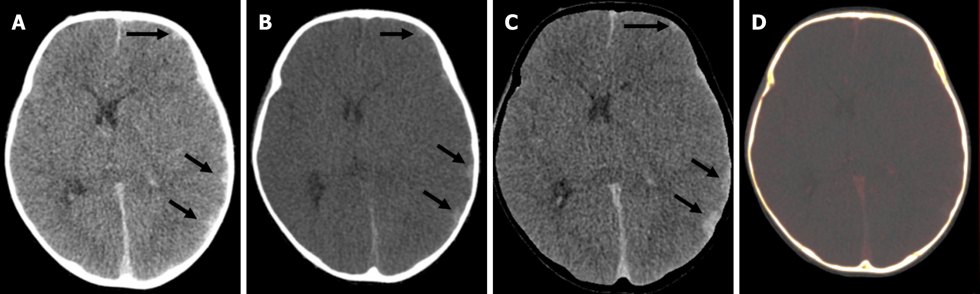
Figure 7 A 4-year-old male with hemophilia A who presented with acute headache and neck rigidity.
A: Axial mixed-enhanced CT head images showed subtle extra-axial high-density lesions (indicated by arrows) in the left frontal and parietal lobes, resulting in a slight midline shift to the opposite side; B: The virtual non-contrast image confirmed that the hyperdensity (arrows) represented an acute bleed and not abnormal enhancement; C: Bone removal tool of dual energy demonstrated the bleed more clearly (arrows); D: Iodine overlay image did not show any iodine uptake in the region of the bleed.
- Citation: Saini S, Bhatia A, Bansal A, Saxena AK, Sodhi KS. Dual-energy computed tomography in children: Technique and clinical applications. World J Clin Pediatr 2025; 14(4): 108823
- URL: https://www.wjgnet.com/2219-2808/full/v14/i4/108823.htm
- DOI: https://dx.doi.org/10.5409/wjcp.v14.i4.108823