
















©The Author(s) 2025.
World J Clin Pediatr. Dec 9, 2025; 14(4): 108329
Published online Dec 9, 2025. doi: 10.5409/wjcp.v14.i4.108329
Published online Dec 9, 2025. doi: 10.5409/wjcp.v14.i4.108329
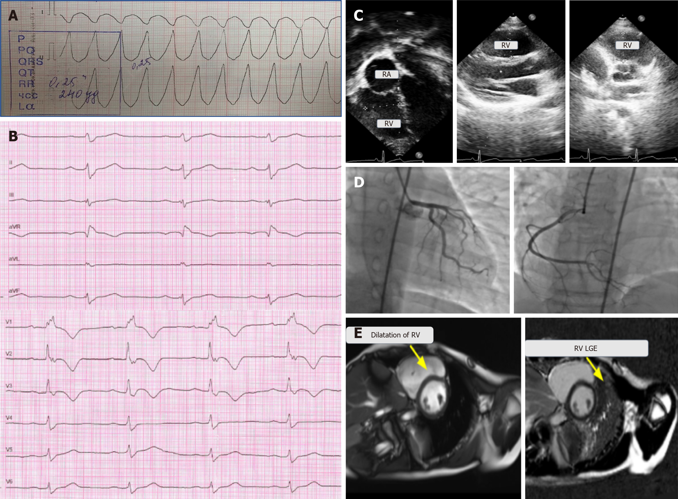
Figure 1 Electrocardiography, echocardiography, angiography, and cardiac magnetic resonance imaging of a 13-year-old boy with a single episode of syncope and ventricular tachycardia.
A: Electrocardiography (ECG) fragment with wide complex QRS tachycardia and a heart rate of 250 per minute; B: ECG during chest pain, sinus rhythm with a heart rate of 64 bpm, and right bundle branch block; C: Echocardiography—right ventricle dilation in the four-chamber position and parasternal position in the long and short axes; D: Type of coronary blood supply—right, coronary arteries with no angiographic signs of atherosclerotic lesions, no local stenosis, and satisfactory blood flow; E: Cardiac magnetic resonance imaging—right ventricular (RV) dilation and late gadolinium enhancement in the RV myocardium. RA: Right atrial; RV: Right ventricular; LGE: Late gadolinium enhancement.
- Citation: Nikitina E, Kofeynikova O, Zlotina A, Pervunina T, Vasichkina E, Golovkin A, Kalinina O, Kostareva A. Arrhythmogenic cardiomyopathy in children, on the link between injurious mutations and inflammation: Two case reports and review of the literature. World J Clin Pediatr 2025; 14(4): 108329
- URL: https://www.wjgnet.com/2219-2808/full/v14/i4/108329.htm
- DOI: https://dx.doi.org/10.5409/wjcp.v14.i4.108329