Copyright
©The Author(s) 2024.
World J Clin Pediatr. Dec 9, 2024; 13(4): 100493
Published online Dec 9, 2024. doi: 10.5409/wjcp.v13.i4.100493
Published online Dec 9, 2024. doi: 10.5409/wjcp.v13.i4.100493
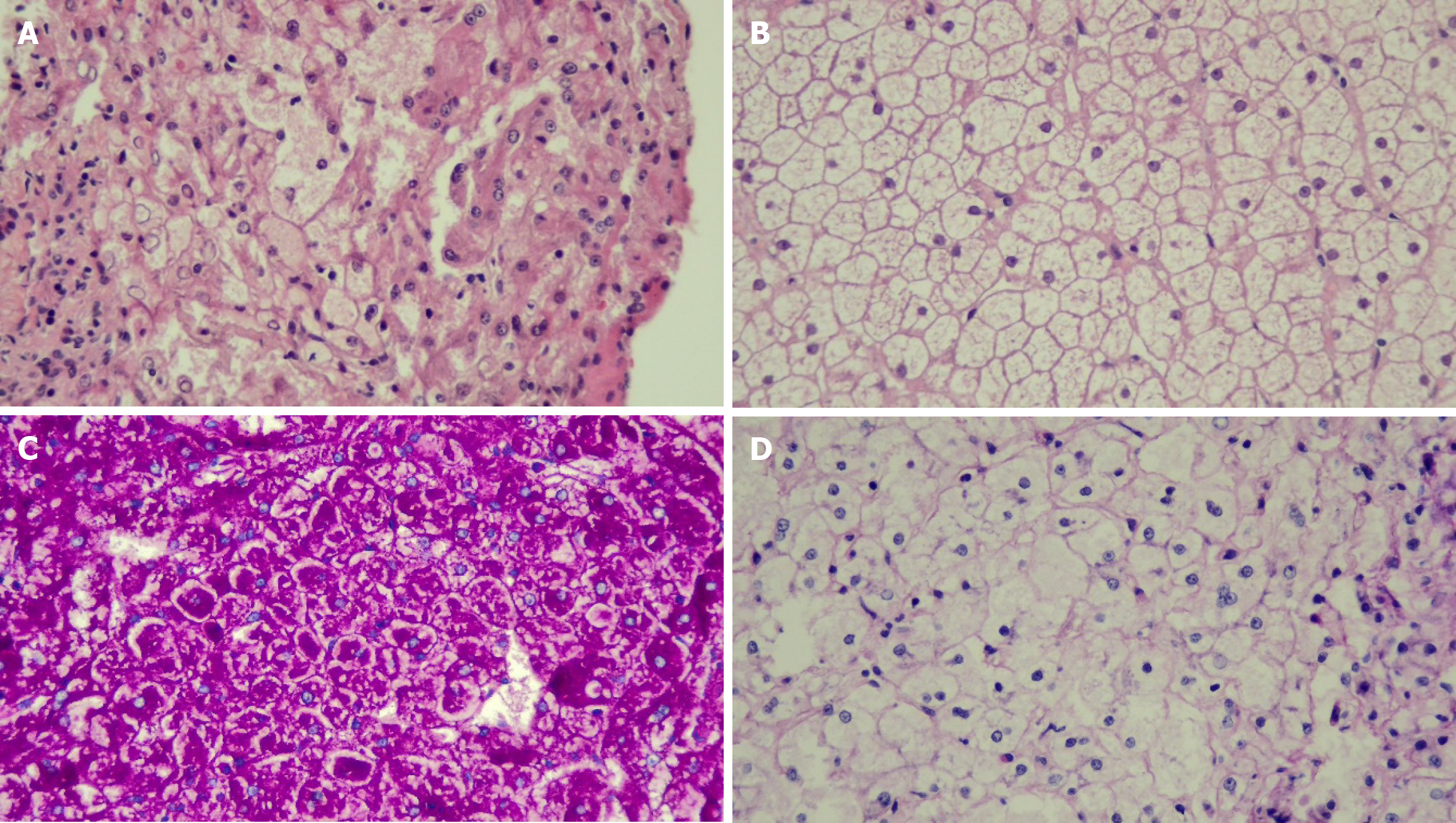
Figure 2 Light microscopy of liver histopathology with high-power magnification (× 40).
A: Patient 6 [glycogen storage disease (GSD) type III] hematoxylin and eosin stain (HE) shows swelling of hepatocytes; B: Patient 7 (GSD type VI) HE stain shows swelling of hepatolcytes with some material deposit inside; C: Patient 7 with periodic acid-Schiff (PAS) staining; D: Patient 7 with PAS-diastase (PAS-D) staining. Hepatocytes were stained with PAS and were mostly digested by PAS-D.
- Citation: Vanduangden J, Ittiwut R, Ittiwut C, Phewplung T, Sanpavat A, Sintusek P, Suphapeetiporn K. Molecular profiles and long-term outcomes of Thai children with hepatic glycogen storage disease in Thailand. World J Clin Pediatr 2024; 13(4): 100493
- URL: https://www.wjgnet.com/2219-2808/full/v13/i4/100493.htm
- DOI: https://dx.doi.org/10.5409/wjcp.v13.i4.100493


