Copyright
©The Author(s) 2023.
World J Clin Pediatr. Jun 9, 2023; 12(3): 97-106
Published online Jun 9, 2023. doi: 10.5409/wjcp.v12.i3.97
Published online Jun 9, 2023. doi: 10.5409/wjcp.v12.i3.97
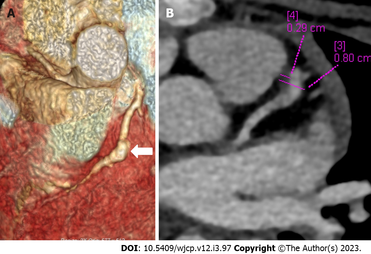
Figure 5 Computed tomography coronary angiography images depicting complications in coronary artery abnormalities (thrombus).
Computed tomography coronary angiography [(A: Volume rendered image; B: Axial image in plane of left anterior descending (LAD)] during follow up at shows a giant fusiform aneurysm in mid segment of LAD (thick arrow in A) with a hypodense plaque like tissue attached to the anterior wall (B) suggestive of thrombus. Child was having chest pain and ECHO at presentation and during the current episode was reported as normal; ECHO fails to elicit coronary artery abnormalities and its complication of thrombus in the aneurysm.
- Citation: Singhal M, Pilania RK, Gupta P, Johnson N, Singh S. Emerging role of computed tomography coronary angiography in evaluation of children with Kawasaki disease. World J Clin Pediatr 2023; 12(3): 97-106
- URL: https://www.wjgnet.com/2219-2808/full/v12/i3/97.htm
- DOI: https://dx.doi.org/10.5409/wjcp.v12.i3.97


