
















©The Author(s) 2023.
World J Clin Pediatr. Jun 9, 2023; 12(3): 97-106
Published online Jun 9, 2023. doi: 10.5409/wjcp.v12.i3.97
Published online Jun 9, 2023. doi: 10.5409/wjcp.v12.i3.97
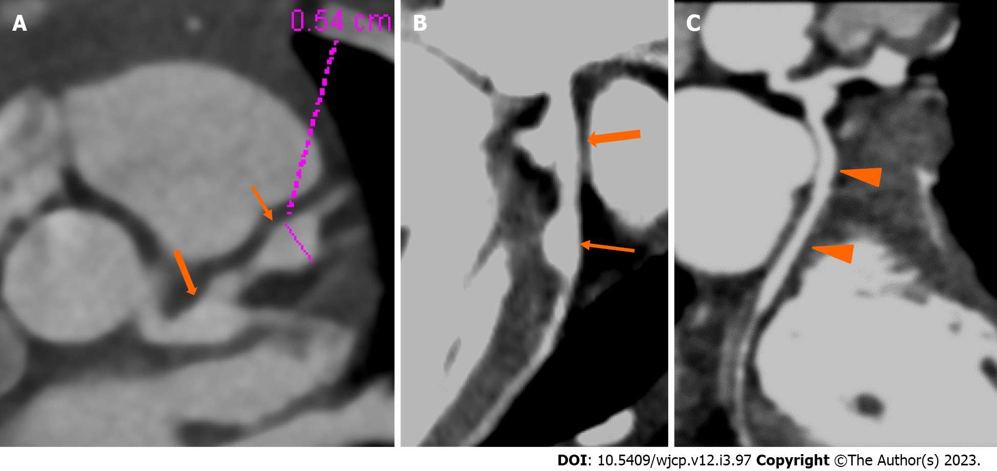
Figure 2 Computed tomography coronary angiography images showing its strength to evaluate coronary artery abnormalities in left circumflex artery.
A: 3 years male child at presentation shows fusiform aneurysm at bifurcation of left main coronary artery [left main coronary artery (LMCA)- thick arrows in A and B] with extension into osteo-proximal segment of left anterior descending (LAD). Note a skip fusiform aneurysm in proximal LAD (thin arrow in b). C: Proximal and mid segments of left circumflex (LCX) are dilated (arrow heads in C). Echo demonstrated LMCA and LAD aneurysms however cannot evaluate LCX due to its orientation and course.
- Citation: Singhal M, Pilania RK, Gupta P, Johnson N, Singh S. Emerging role of computed tomography coronary angiography in evaluation of children with Kawasaki disease. World J Clin Pediatr 2023; 12(3): 97-106
- URL: https://www.wjgnet.com/2219-2808/full/v12/i3/97.htm
- DOI: https://dx.doi.org/10.5409/wjcp.v12.i3.97