
















©The Author(s) 2022.
World J Clin Pediatr. May 9, 2022; 11(3): 270-288
Published online May 9, 2022. doi: 10.5409/wjcp.v11.i3.270
Published online May 9, 2022. doi: 10.5409/wjcp.v11.i3.270
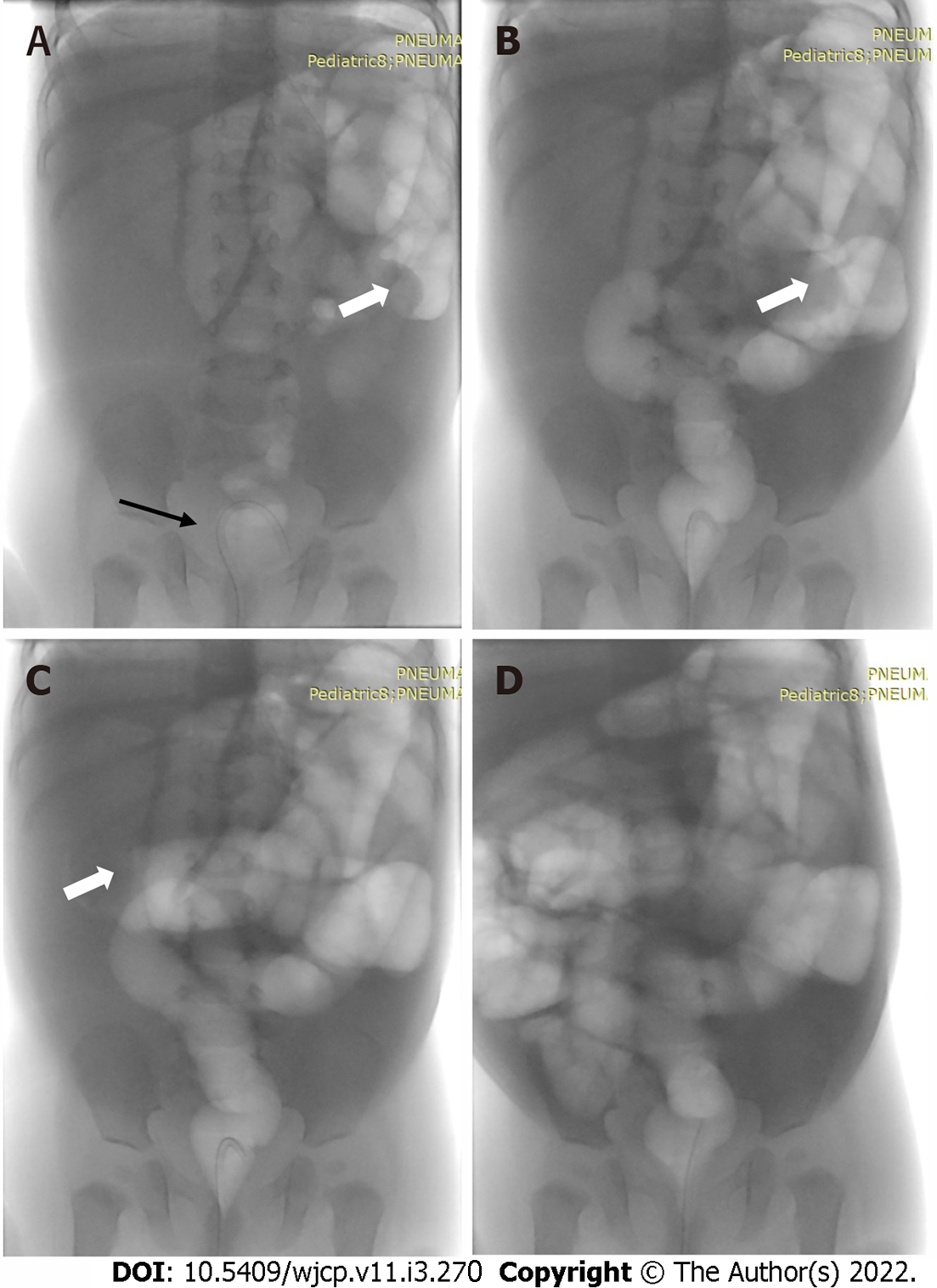
Figure 22 Pneumatic reduction of intussusception under fluoroscopy.
A: The patient is positioned supine with a feeding tube within the rectum (black arrow); B and C: The intussusceptum is seen in the left hypochondrium (open arrow) given by the meniscus sign; subsequent spots after air inflation show that the intussusceptum has moved proximally, as well as reflux of air in the small bowel after successful reduction (D) of the intussusception.
- Citation: Chandel K, Jain R, Bhatia A, Saxena AK, Sodhi KS. Bleeding per rectum in pediatric population: A pictorial review. World J Clin Pediatr 2022; 11(3): 270-288
- URL: https://www.wjgnet.com/2219-2808/full/v11/i3/270.htm
- DOI: https://dx.doi.org/10.5409/wjcp.v11.i3.270