Copyright
©The Author(s) 2021.
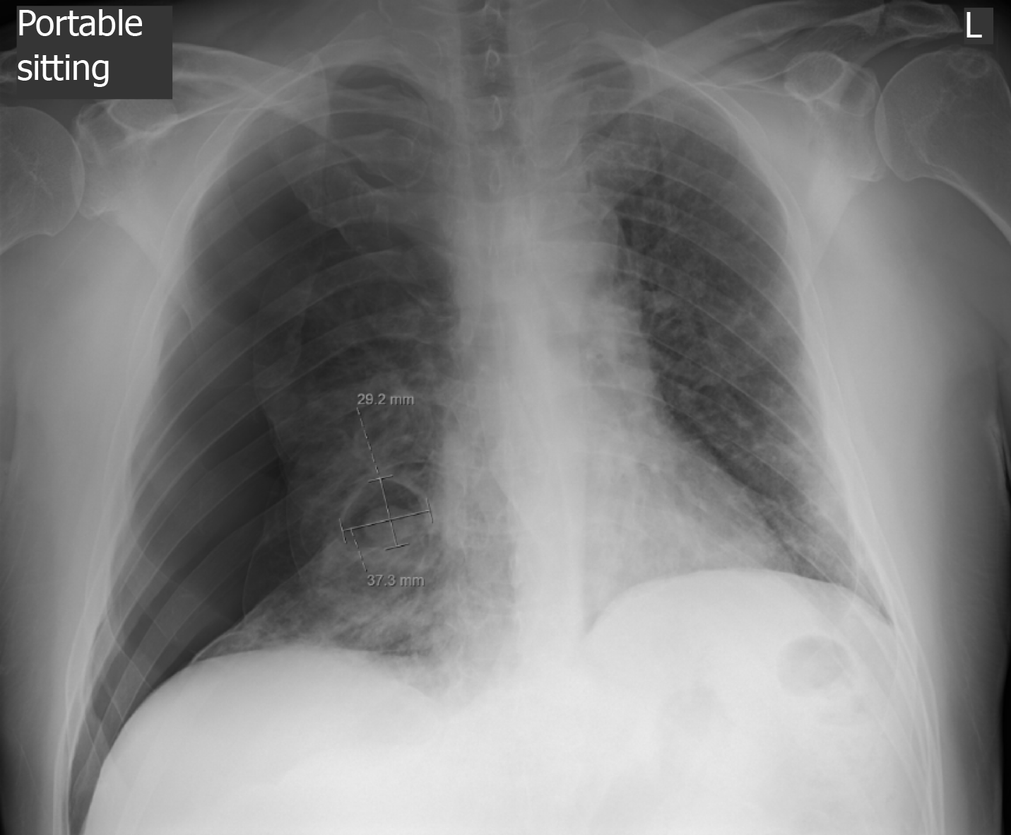
Figure 1 Chest radiograph of the patient.
The image shows development of large right-sided pneumothorax with associated collapse of the right lung, as well as a 3.7 cm × 2.9 cm air-filled cystic structure with a round solid dependent component in the right lower lung.
- Citation: Mathew J, Cherukuri SV, Dihowm F. SARS-CoV-2 with concurrent coccidioidomycosis complicated by refractory pneumothorax in a Hispanic male: A case report and literature review. World J Respirol 2021; 11(1): 1-11
- URL: https://www.wjgnet.com/2218-6255/full/v11/i1/1.htm
- DOI: https://dx.doi.org/10.5320/wjr.v11.i1.1


