Copyright
©The Author(s) 2017.
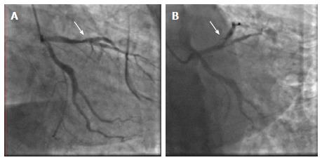
Figure 1 Left coronary angiogram.
Preintervention angiogram of the left anterior descending artery revealed the presence of 70% calcified stenosis of its middle part (A), involving the bifurcation of the first diagonal branch, with a calcific stenosis at its middle portion (B). Arrow indicates the site of the lesions.
- Citation: Carbone A, Formisano T, Natale F, Cappelli Bigazzi M, Tartaglione D, Golia E, Gragnano F, Crisci M, Bianchi RM, Calabrò R, Russo MG, Calabrò P. Management of unstable angina in a patient with Haemophilia A. World J Hematol 2017; 6(2): 28-31
- URL: https://www.wjgnet.com/2218-6204/full/v6/i2/28.htm
- DOI: https://dx.doi.org/10.5315/wjh.v6.i2.28


