Copyright
©2014 Baishideng Publishing Group Inc.
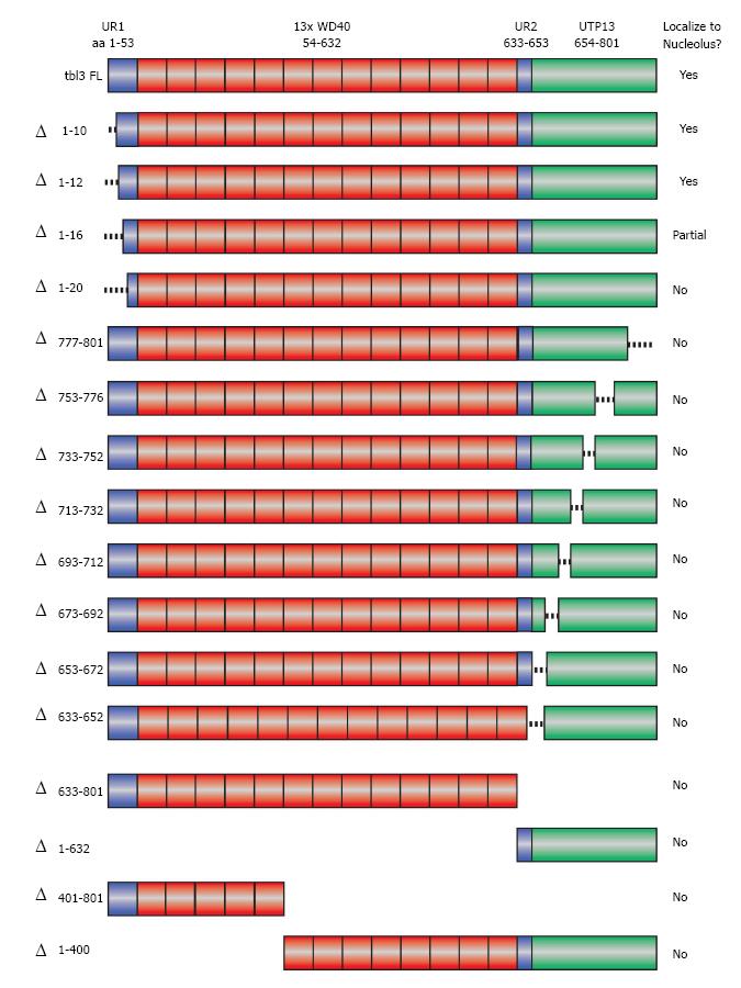
Figure 4 Scanning deletions of tbl3 and their effects on nucleolar targeting.
A schematic summary of the deletions of transducin β-like 3 (tbl3) tested in the localization study. Shown at the very top is the modular structure of tbl3. “Δ” indicates deleted aa. Deletion mutants were expressed as C-terminal enhanced green fluorescent protein (EGFP) fusion proteins in NIH3T3 and examined by fluorescence microscopy. The results of localization are summarized in the right column. UR1: Unique region 1 (aa 1-53); WD40: Region containing thirteen WD40 repeats (aa 54-632); UR2: Unique region 2 (aa 633-653); UTP13: Conserved C-terminal domain (aa 654-801).
-
Citation: Wang J, Tsai S.
Tbl3 encodes a WD40 nucleolar protein with regulatory roles in ribosome biogenesis. World J Hematol 2014; 3(3): 93-104 - URL: https://www.wjgnet.com/2218-6204/full/v3/i3/93.htm
- DOI: https://dx.doi.org/10.5315/wjh.v3.i3.93


