Copyright
©The Author(s) 2023.
World J Hematol. Dec 29, 2023; 10(4): 42-47
Published online Dec 29, 2023. doi: 10.5315/wjh.v10.i4.42
Published online Dec 29, 2023. doi: 10.5315/wjh.v10.i4.42
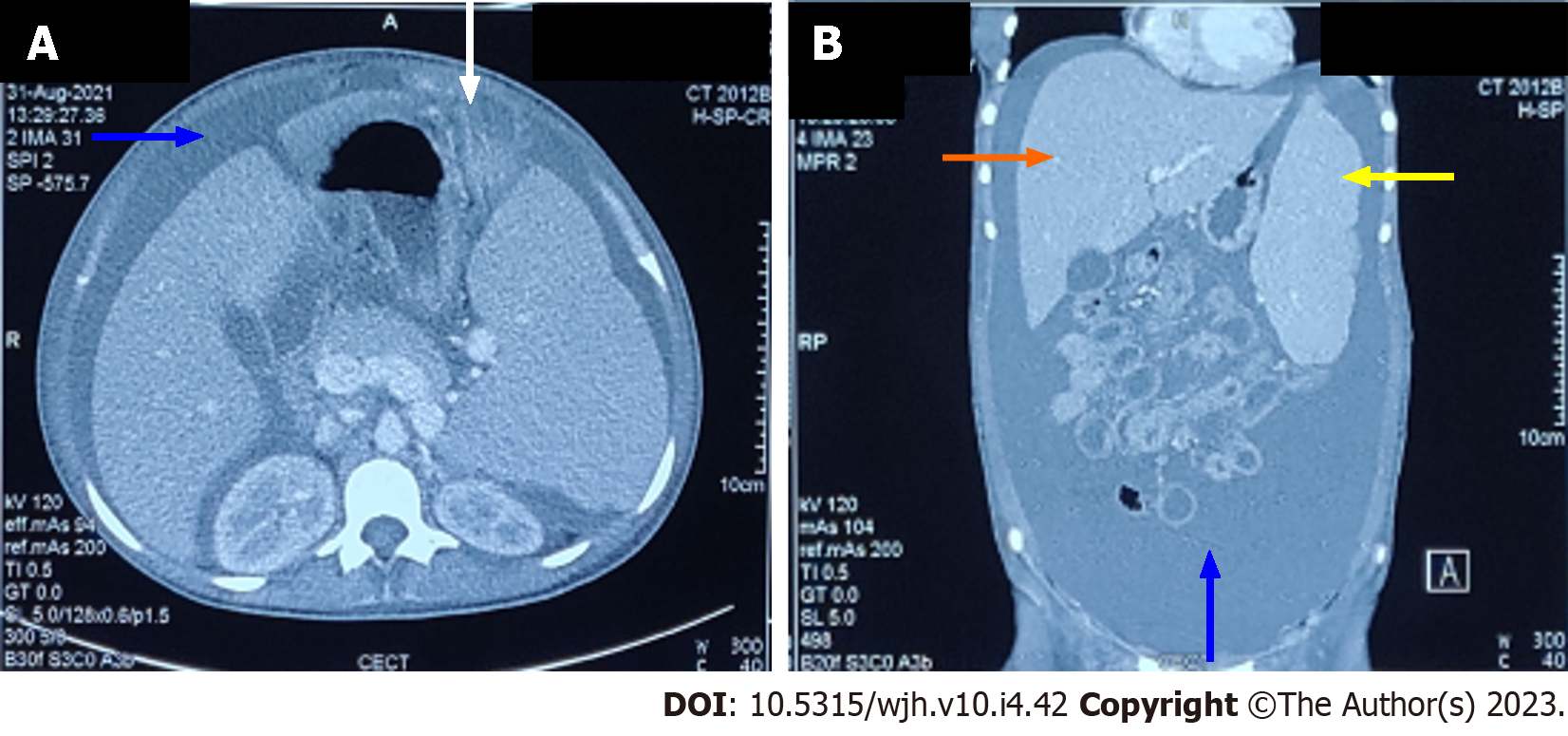
Figure 2 Contrast enhanced computed tomography of abdomen showing ascites (blue arrow), omental caking (white arrow), hepatomegaly (orange arrow), and splenomegaly (yellow arrow).
A: Axial section; B: Coronal section.
- Citation: Mishra R, Garg S, Bharti P, Malla DR, Rohatgi I, Gautam S. Unusual presentation of extramedullary blast crisis in chronic myeloid leukemia: A case report. World J Hematol 2023; 10(4): 42-47
- URL: https://www.wjgnet.com/2218-6204/full/v10/i4/42.htm
- DOI: https://dx.doi.org/10.5315/wjh.v10.i4.42


