Copyright
©The Author(s) 2015.
World J Anesthesiol. Jul 27, 2015; 4(2): 44-48
Published online Jul 27, 2015. doi: 10.5313/wja.v4.i2.44
Published online Jul 27, 2015. doi: 10.5313/wja.v4.i2.44
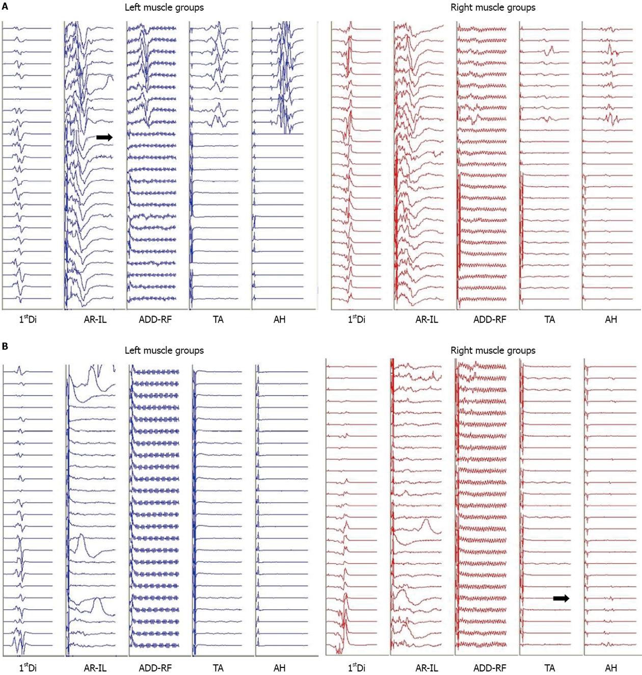
Figure 2 Motor evoked potentials during vertebral column resection (A) and at surgical closure (B).
A: Abrupt loss of the left and right lower extremity motor evoked potentials (MEPs) during the vertebral column resection (dark arrows). Transcranial electric stimulation is delivered between two subdermal needle electrodes inserted 2 cm anterior to C1-C2 (International 10-20 System) overlying the motor cortex region. Trains of 5 to 9 pulses, spaced at an interstimulus interval ranging from 1.1 to 4.1 ms, are delivered with constant voltage (200-500 V) at the anode. Resultant compound muscle action potentials are recorded using subdermal needle electrodes, in a bipolar montage. These myogenic responses are recorded bilaterally from the first dorsal interosseous muscles (1stDi) in the upper extremity and lower extremity MEPs are recorded from the left and right abdominal rectus-iliopsoas (AR-IL), adductors-rectus femoris (ADD-RF), tibialis anterior (TA), and abductor hallucis (AH) muscles. Muscle groups are linked on occasion in order to maximize nerve root coverage. These unaveraged compound muscle action potentials are recorded through a 30-1000 Hz bandpass filter and are displayed in a 100 ms window; B: Closing left and right MEP responses. The dark arrow indicates the onset of a very small recovery of the right AH. There are no responses present from the left lower extremity muscle groups. Upon emergence from anesthesia the patient was noted to be purposefully moving all but the left lower limb coinciding with his MEP responses. Intermittent responses recorded from the left and right AR-IL were a result of movement artifact. Stimulation and recording parameters are similar to Figure 2A.
- Citation: Karsli C, Strantzas S, Finnerty O, Holmes L, Lewis S. Bradycardia and hypotension during pediatric scoliosis surgery-hypovolemia or spinal shock? World J Anesthesiol 2015; 4(2): 44-48
- URL: https://www.wjgnet.com/2218-6182/full/v4/i2/44.htm
- DOI: https://dx.doi.org/10.5313/wja.v4.i2.44


