Copyright
©The Author(s) 2018.
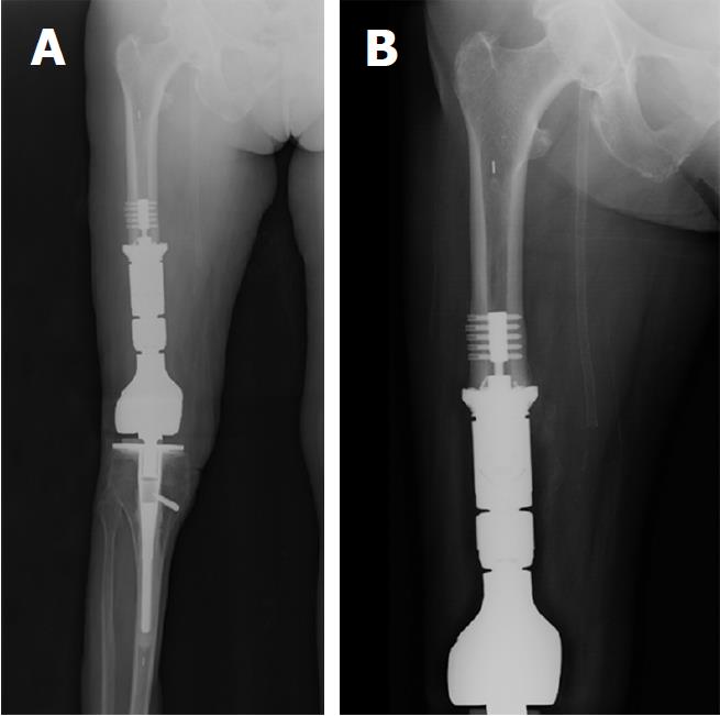
Figure 3 Postoperative radiographs showing the additional bone resection, resection of the soft tissue pseudotumor, and the definitive reconstruction with an extended femoral endoprosthesis.
A: Four week post-operative radiograph showing resection of the distal 9 cm of femoral diaphysis and reconstruction with a 9 cm intercalary segment. The intercalary segment is secured to the femoral diaphysis with a femoral compress device (Zimmer Biomet, Warsaw, IN, United States); B: One-year postoperative radiograph showing distal femoral bone remodeling and bone integration into the porous end of the femoral compress device. The patient’s thigh pain was completely resolved and her gait returned to a reciprocating smooth gait that appeared symmetrical to the contralateral leg.
- Citation: Chowdhry M, Dipane MV, McPherson EJ. Periosteal pseudotumor in complex total knee arthroplasty resembling a neoplastic process. World J Orthop 2018; 9(5): 72-77
- URL: https://www.wjgnet.com/2218-5836/full/v9/i5/72.htm
- DOI: https://dx.doi.org/10.5312/wjo.v9.i5.72


