Copyright
©The Author(s) 2017.
World J Orthop. Sep 18, 2017; 8(9): 688-696
Published online Sep 18, 2017. doi: 10.5312/wjo.v8.i9.688
Published online Sep 18, 2017. doi: 10.5312/wjo.v8.i9.688
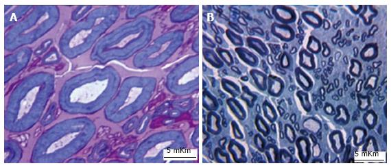
Figure 3 Fragments of canine Tn semithin sections, WA30.
A: Some large myelinated nerve fibres has visibly thinned axons and thickened myelin sheaths, М-group, methylene blue - basic fuchsin stain, magnification 1250 ×; B: Two large nerve fibers (in the lower part of the image) are hypomyelinated; А-group, toluidine blue stain, magnification 500 ×.
- Citation: Shchudlo N, Varsegova T, Stupina T, Shchudlo M, Saifutdinov M, Yemanov A. Benefits of Ilizarov automated bone distraction for nerves and articular cartilage in experimental leg lengthening. World J Orthop 2017; 8(9): 688-696
- URL: https://www.wjgnet.com/2218-5836/full/v8/i9/688.htm
- DOI: https://dx.doi.org/10.5312/wjo.v8.i9.688


