Copyright
©The Author(s) 2017.
World J Orthop. Sep 18, 2017; 8(9): 660-673
Published online Sep 18, 2017. doi: 10.5312/wjo.v8.i9.660
Published online Sep 18, 2017. doi: 10.5312/wjo.v8.i9.660
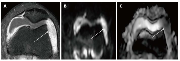
Figure 6 Septic synovitis.
Magnetic resonance imaging findings in a 22-year-old man with knee pain and fever. A: Axial post contrast fat-suppressed TSE T1-weighted image shows a large joint effusion with synovial thickening and intense enhancement; B, C: DWI with a b value of 800 s/mm2 and corresponding ADC map demonstrate areas of severely restricted diffusion (ADC: 1, 8 × 10⁻³ mm2/s) within the articular fluid at the lateral patellar recess consistent with exudate, as proven by arthrocentesis. DWI: Diffusion-weighted imaging; ADC: Apparent diffusion coefficient; TSE: Turbo spin echo.
- Citation: Martín Noguerol T, Luna A, Gómez Cabrera M, Riofrio AD. Clinical applications of advanced magnetic resonance imaging techniques for arthritis evaluation. World J Orthop 2017; 8(9): 660-673
- URL: https://www.wjgnet.com/2218-5836/full/v8/i9/660.htm
- DOI: https://dx.doi.org/10.5312/wjo.v8.i9.660


