
















©The Author(s) 2017.
World J Orthop. Sep 18, 2017; 8(9): 660-673
Published online Sep 18, 2017. doi: 10.5312/wjo.v8.i9.660
Published online Sep 18, 2017. doi: 10.5312/wjo.v8.i9.660
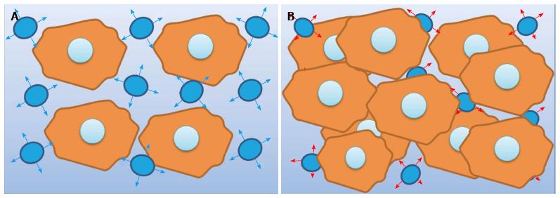
Figure 2 Diffusion-weighted imaging.
A: Free water diffusion. The diagram represents the free motion of water molecules in the extracellular space between cells in normal tissue; B: Restricted water diffusion. The diagram represents the restricted motion of water molecules in the extracellular space due to hypercellularity. Another condition that leads to a decrease in the extracellular space is the presence of cytotoxic edema, while the presence of debris and detritus as in the case of abscesses may result also in restricted diffusion.
- Citation: Martín Noguerol T, Luna A, Gómez Cabrera M, Riofrio AD. Clinical applications of advanced magnetic resonance imaging techniques for arthritis evaluation. World J Orthop 2017; 8(9): 660-673
- URL: https://www.wjgnet.com/2218-5836/full/v8/i9/660.htm
- DOI: https://dx.doi.org/10.5312/wjo.v8.i9.660