Copyright
©The Author(s) 2017.
World J Orthop. Feb 18, 2017; 8(2): 163-169
Published online Feb 18, 2017. doi: 10.5312/wjo.v8.i2.163
Published online Feb 18, 2017. doi: 10.5312/wjo.v8.i2.163
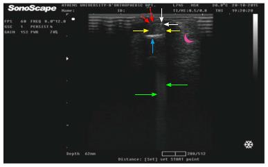
Figure 4 In order to be assured for the right position of the knife, we are transferred in transverse view of the tendons (flexor’s transverse cut: Yellow arrows, lying on the bone: Light blue arrow).
Here, we certify that the tip of the knife (tip as a white dot: white arrows, sending its characteristic acoustic shadow: Green arrows) is attaching the volar end of the tendons (without penetrating them), under the A1 pulley (the sheath is appeared as a thin line under the red arrows). Moreover, it is vital to avoid the neurovascular digital structures (digital artery in the curved side of the purple moon).
- Citation: Nikolaou VS, Malahias MA, Kaseta MK, Sourlas I, Babis GC. Comparative clinical study of ultrasound-guided A1 pulley release vs open surgical intervention in the treatment of trigger finger. World J Orthop 2017; 8(2): 163-169
- URL: https://www.wjgnet.com/2218-5836/full/v8/i2/163.htm
- DOI: https://dx.doi.org/10.5312/wjo.v8.i2.163


