Copyright
©The Author(s) 2017.
World J Orthop. Feb 18, 2017; 8(2): 156-162
Published online Feb 18, 2017. doi: 10.5312/wjo.v8.i2.156
Published online Feb 18, 2017. doi: 10.5312/wjo.v8.i2.156
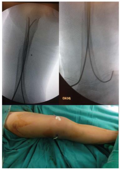
Figure 2 Intraoperative X-rays showing the correct positioning of titanium elastic nail.
Entry points were performed, almost 2.5 cm proximal to the distal physis, one medial and one lateral. To facilitate the removal of the titanium elastic nail, its tail could be left over the skin surface as evident from the clinical intraoperative picture.
- Citation: Donati F, Mazzitelli G, Lillo M, Menghi A, Conti C, Valassina A, Marzetti E, Maccauro G. Titanium elastic nailing in diaphyseal femoral fractures of children below six years of age. World J Orthop 2017; 8(2): 156-162
- URL: https://www.wjgnet.com/2218-5836/full/v8/i2/156.htm
- DOI: https://dx.doi.org/10.5312/wjo.v8.i2.156


