Copyright
©The Author(s) 2017.
World J Orthop. Dec 18, 2017; 8(12): 935-945
Published online Dec 18, 2017. doi: 10.5312/wjo.v8.i12.935
Published online Dec 18, 2017. doi: 10.5312/wjo.v8.i12.935
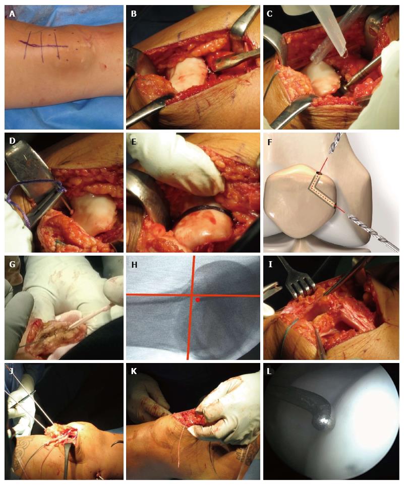
Figure 2 Step-by-step description of the combined surgical procedure.
A: Short skin incision running from the medial part of the patella to the distal part of the quadriceps tendon; B: Using a curved osteotome, a thin osteochondral flap is chiselled off the trochlea; C: Trochlear bone is deepened using chisels and a high-speed burr; D: Vicryl band is passed from the distal beginning of the formed groove through the condyle using a curved needle; E: Starting at the junction between the trochlear cartilage and the anterior femoral bone, the other end of the band is passed through the femur. After pressing the flap onto the bone, the band is fastened; F: Drilling of a V-shaped tunnel coming from the superomedial pole and from the middle of the medial facet of the patella; G: Gracilis tendon is passed through the drill holes; H: Femoral medial patellofemoral ligament (MPFL) insertion side is marked with a K wire under fluoroscopic guidance; I: Prepared soft tissue tunnel between both layers of the medial retinaculum is running down to the femoral MPFL insertion; J: Two divergent beath pins are passed from the blind ending tunnel through the lateral femoral cortex; K: After passing the strands to the lateral femur, a temporary knot is tied down in approximately 30° knee flexion; L: Arthroscopy of the trochlear groove after trochleoplasty.
- Citation: von Engelhardt LV, Weskamp P, Lahner M, Spahn G, Jerosch J. Deepening trochleoplasty combined with balanced medial patellofemoral ligament reconstruction for an adequate graft tensioning. World J Orthop 2017; 8(12): 935-945
- URL: https://www.wjgnet.com/2218-5836/full/v8/i12/935.htm
- DOI: https://dx.doi.org/10.5312/wjo.v8.i12.935


