
















©The Author(s) 2017.
World J Orthop. Oct 18, 2017; 8(10): 747-753
Published online Oct 18, 2017. doi: 10.5312/wjo.v8.i10.747
Published online Oct 18, 2017. doi: 10.5312/wjo.v8.i10.747
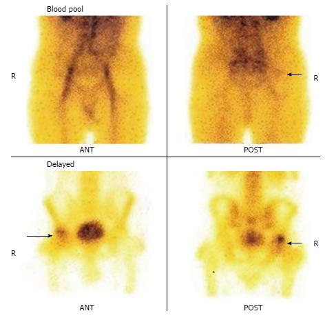
Figure 5 Blood pool images of the patient (as mentioned Figure 4) show minimally increased tracer uptake in the region of right hip.
The delayed images show increased tracer uptake in the right hip region with no definite photopenic area.
- Citation: Agrawal K, Tripathy SK, Sen RK, Santhosh S, Bhattacharya A. Nuclear medicine imaging in osteonecrosis of hip: Old and current concepts. World J Orthop 2017; 8(10): 747-753
- URL: https://www.wjgnet.com/2218-5836/full/v8/i10/747.htm
- DOI: https://dx.doi.org/10.5312/wjo.v8.i10.747