Copyright
©The Author(s) 2016.
World J Orthop. Jul 18, 2016; 7(7): 406-417
Published online Jul 18, 2016. doi: 10.5312/wjo.v7.i7.406
Published online Jul 18, 2016. doi: 10.5312/wjo.v7.i7.406
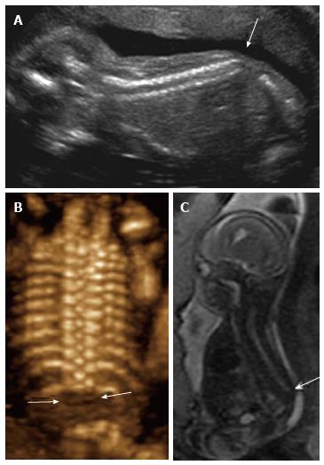
Figure 11 Sacral agenesis.
2D sagittal grayscale ultrasound (A), coronal 3D ultrasound (B), and parasagittal T1 weighted image (C) of a 19 wk fetus demonstrate absence of the fetal spine caudal to T12 (arrows), consistent with sacral agenesis/caudal regression syndrome. The mother was known to have type 1 diabetes, a risk factor for this condition.
- Citation: Upasani VV, Ketwaroo PD, Estroff JA, Warf BC, Emans JB, Glotzbecker MP. Prenatal diagnosis and assessment of congenital spinal anomalies: Review for prenatal counseling. World J Orthop 2016; 7(7): 406-417
- URL: https://www.wjgnet.com/2218-5836/full/v7/i7/406.htm
- DOI: https://dx.doi.org/10.5312/wjo.v7.i7.406


