
















©The Author(s) 2016.
World J Orthop. May 18, 2016; 7(5): 272-279
Published online May 18, 2016. doi: 10.5312/wjo.v7.i5.272
Published online May 18, 2016. doi: 10.5312/wjo.v7.i5.272
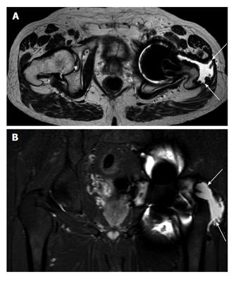
Figure 3 Axial (A) and coronal (B) magnetic resonance imaging.
Example of abductor stripping secondary to a pseudotumour (marked by arrows). The pseudotumour can be seen traversing the posterior hip around the greater tuberosity onto its lateral aspect, which is now void of abductor tendon insertion.
- Citation: Berber R, Skinner JA, Hart AJ. Management of metal-on-metal hip implant patients: Who, when and how to revise? World J Orthop 2016; 7(5): 272-279
- URL: https://www.wjgnet.com/2218-5836/full/v7/i5/272.htm
- DOI: https://dx.doi.org/10.5312/wjo.v7.i5.272