Copyright
©The Author(s) 2016.
World J Orthop. Dec 18, 2016; 7(12): 843-846
Published online Dec 18, 2016. doi: 10.5312/wjo.v7.i12.843
Published online Dec 18, 2016. doi: 10.5312/wjo.v7.i12.843
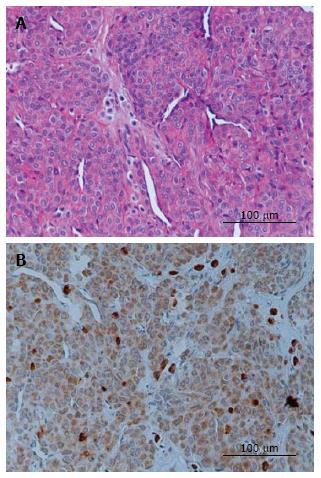
Figure 1 Histopathologic examination.
A: The tumor is composed of multiple vascular channels lined by endothelial cells and aggregates of round cells with darkly staining round to ovoid nuclei and eosinophilic cytoplasm (hematoxylin and eosin stain, × 400); B: The tumour cells are strongly positive for vascular endothelial growth factor (VEGF stain, × 400). Strong cytoplasmic staining for VEGF in the tumor parenchyma. VEGF: Vascular endothelial growth factor.
- Citation: Honsawek S, Kitidumrongsook P, Luangjarmekorn P, Pataradool K, Thanakit V, Patradul A. Glomus tumors of the fingers: Expression of vascular endothelial growth factor. World J Orthop 2016; 7(12): 843-846
- URL: https://www.wjgnet.com/2218-5836/full/v7/i12/843.htm
- DOI: https://dx.doi.org/10.5312/wjo.v7.i12.843


