
















©The Author(s) 2016.
World J Orthop. Dec 18, 2016; 7(12): 785-792
Published online Dec 18, 2016. doi: 10.5312/wjo.v7.i12.785
Published online Dec 18, 2016. doi: 10.5312/wjo.v7.i12.785
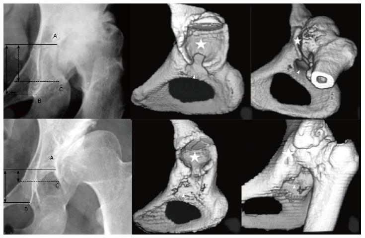
Figure 3 Images illustrating the two subtypes of low dislocation: B1 and B2.
Three points must be recognised on radiographs: (A) the superior limit of the true acetabulum; (B) the inferior point of the teardrop; (C) the most inferior point of the false acetabulum. Three dimensional-computed tomography scans may help to determine the superior limit of the true acetabulum, when it is not clear in plain radiographs. Asterisks depict false acetabulum and arrowheads true acetabulum.
- Citation: Hartofilakidis G, Lampropoulou-Adamidou K. Lessons learned from study of congenital hip disease in adults. World J Orthop 2016; 7(12): 785-792
- URL: https://www.wjgnet.com/2218-5836/full/v7/i12/785.htm
- DOI: https://dx.doi.org/10.5312/wjo.v7.i12.785