Copyright
©The Author(s) 2016.
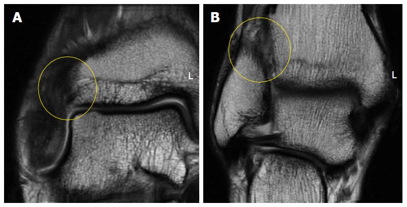
Figure 2 Coronal fast-spin echo proton density magnetic resonance images of the right ankle seen in Figure 1.
Disruption and remodeling of the anterior inferior tibiofibular ligament (A; yellow circle) and interosseous ligament (B; yellow circle) can be appreciated.
- Citation: Walls RJ, Ross KA, Fraser EJ, Hodgkins CW, Smyth NA, Egan CJ, Calder J, Kennedy JG. Football injuries of the ankle: A review of injury mechanisms, diagnosis and management. World J Orthop 2016; 7(1): 8-19
- URL: https://www.wjgnet.com/2218-5836/full/v7/i1/8.htm
- DOI: https://dx.doi.org/10.5312/wjo.v7.i1.8


