Copyright
©The Author(s) 2015.
World J Orthop. Oct 18, 2015; 6(9): 688-704
Published online Oct 18, 2015. doi: 10.5312/wjo.v6.i9.688
Published online Oct 18, 2015. doi: 10.5312/wjo.v6.i9.688
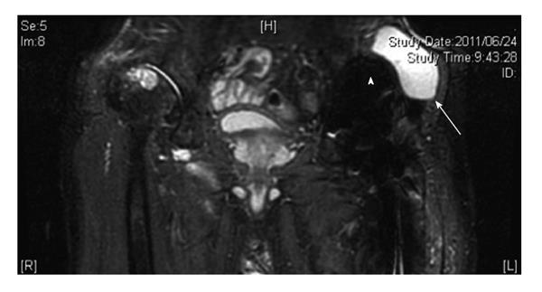
Figure 5 A 76-year-old male with a pseudotumor following cemented total hip arthroplasty, which was implanted 18 years earlier.
Coronal short tau inversion recovery magnetic resonance image shows a cystic lesion (arrow) adjacent to the acetabular component (arrowhead).
- Citation: Yukata K, Nakai S, Goto T, Ikeda Y, Shimaoka Y, Yamanaka I, Sairyo K, Hamawaki JI. Cystic lesion around the hip joint. World J Orthop 2015; 6(9): 688-704
- URL: https://www.wjgnet.com/2218-5836/full/v6/i9/688.htm
- DOI: https://dx.doi.org/10.5312/wjo.v6.i9.688


