Copyright
©The Author(s) 2015.
World J Orthop. Oct 18, 2015; 6(9): 660-671
Published online Oct 18, 2015. doi: 10.5312/wjo.v6.i9.660
Published online Oct 18, 2015. doi: 10.5312/wjo.v6.i9.660
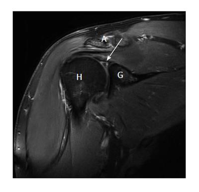
Figure 3 Superior labral anterior posterior III lesion: Findings in direct magnetic resonance arthrography.
The coronal T1-weighted (cor t1 tse fs) fat-saturated image shows the separated triangle (arrow) of the bucket-handle without instability of the labro-bicipital complex. A: Acromion; H: Humeral head; G: Glenoid.
- Citation: Popp D, Schöffl V. Superior labral anterior posterior lesions of the shoulder: Current diagnostic and therapeutic standards. World J Orthop 2015; 6(9): 660-671
- URL: https://www.wjgnet.com/2218-5836/full/v6/i9/660.htm
- DOI: https://dx.doi.org/10.5312/wjo.v6.i9.660


