Copyright
©The Author(s) 2015.
World J Orthop. Mar 18, 2015; 6(2): 202-210
Published online Mar 18, 2015. doi: 10.5312/wjo.v6.i2.202
Published online Mar 18, 2015. doi: 10.5312/wjo.v6.i2.202
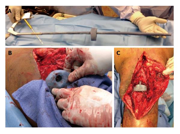
Figure 6 Intraoperative photographs of a temporary arthrodesis for an infected total knee arthroplasty.
A: Photograph shows the long, antibiotic cement-coated intramedullary fusion nail and antibiotic cement-coated spacer prior to insertion. The static antibiotic cement-coated spacer was made with a hole placed centrally that was large enough to accommodate the antibiotic cement-coated nail; B: The nail was inserted to the level of the distal femur, was guided through the central hole in the static spacer, and was guided into the tibia. As the nail was inserted into the tibia, the tibia was held in proper alignment and rotation. When the nail reached the midshaft of the tibia, axial load was applied to the lower extremity to prevent distraction from occurring at the knee; C: The knee wound following spacer insertion and locking of the rod (reprinted with permission from the Rubin Institute for Advanced Orthopedics, Sinai Hospital of Baltimore).
- Citation: Wood JH, Conway JD. Advanced concepts in knee arthrodesis. World J Orthop 2015; 6(2): 202-210
- URL: https://www.wjgnet.com/2218-5836/full/v6/i2/202.htm
- DOI: https://dx.doi.org/10.5312/wjo.v6.i2.202


