Copyright
©The Author(s) 2015.
World J Orthop. Mar 18, 2015; 6(2): 202-210
Published online Mar 18, 2015. doi: 10.5312/wjo.v6.i2.202
Published online Mar 18, 2015. doi: 10.5312/wjo.v6.i2.202
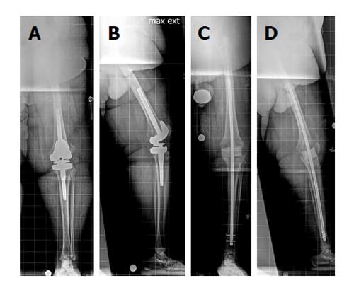
Figure 5 Temporary knee fusion is accomplished by inserting both an antibiotic cement-coated intramedullary knee fusion nail.
Anteroposterior (A) and lateral (B) view radiographs of the left leg in a 71-year-old patient with an infected revision total knee arthroplasty. The patient was morbidly obese. A knee joint gap greater than 5 cm was expected after removal of the infected implants. Knee flexion contracture was also present (B). Temporary knee fusion was planned. Anteroposterior (C) and lateral (D) view radiographs of the left leg obtained postoperatively show the temporary knee fusion, including a long, antibiotic cement-coated intramedullary nail and a static antibiotic cement-coated spacer to fill the knee joint gap (reprinted with permission from the Rubin Institute for Advanced Orthopedics, Sinai Hospital of Baltimore).
- Citation: Wood JH, Conway JD. Advanced concepts in knee arthrodesis. World J Orthop 2015; 6(2): 202-210
- URL: https://www.wjgnet.com/2218-5836/full/v6/i2/202.htm
- DOI: https://dx.doi.org/10.5312/wjo.v6.i2.202


