Copyright
©The Author(s) 2015.
World J Orthop. Mar 18, 2015; 6(2): 202-210
Published online Mar 18, 2015. doi: 10.5312/wjo.v6.i2.202
Published online Mar 18, 2015. doi: 10.5312/wjo.v6.i2.202
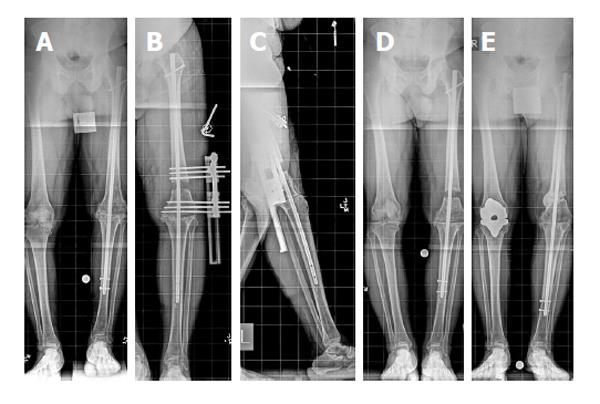
Figure 3 Knee arthrodesis.
A: Anteroposterior view full length standing radiograph shows that the left limb has a 3-cm limb length discrepancy. Note the lift that is used under the left limb; B: Anteroposterior view full length standing radiograph shows the distal femoral osteotomy and lengthening over the existing knee fusion nail. Note that the distal interlocking screws are removed from the nail; C: Full length lateral view radiograph shows that the pins of the external fixator have been placed away from the intramedullary nail; D: Postoperative anteroposterior view full length standing radiograph shows that interlocking screws have been placed and the external fixator has been removed. Note the resolution of the limb length discrepancy; E: Anteroposterior view full length standing radiograph obtained 2 years after lengthening (reprinted with permission from the Rubin Institute for Advanced Orthopedics, Sinai Hospital of Baltimore).
- Citation: Wood JH, Conway JD. Advanced concepts in knee arthrodesis. World J Orthop 2015; 6(2): 202-210
- URL: https://www.wjgnet.com/2218-5836/full/v6/i2/202.htm
- DOI: https://dx.doi.org/10.5312/wjo.v6.i2.202


