Copyright
©The Author(s) 2015.
World J Orthop. Dec 18, 2015; 6(11): 961-969
Published online Dec 18, 2015. doi: 10.5312/wjo.v6.i11.961
Published online Dec 18, 2015. doi: 10.5312/wjo.v6.i11.961
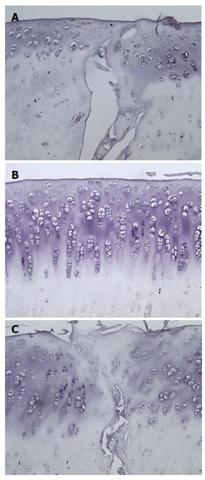
Figure 4 Sagittal sections of an osteochondral graft treated with saline 12 wk following surgery (400 ×).
Transforming growth factor-beta1 antibody staining is positive in cells that appear brown. A: Left interface between the graft and host cartilage; B: Central aspect of the graft; C: Right interface between the graft and host cartilage.
- Citation: Boakye LA, Ross KA, Pinski JM, Smyth NA, Haleem AM, Hannon CP, Fortier LA, Kennedy JG. Platelet-rich plasma increases transforming growth factor-beta1 expression at graft-host interface following autologous osteochondral transplantation in a rabbit model. World J Orthop 2015; 6(11): 961-969
- URL: https://www.wjgnet.com/2218-5836/full/v6/i11/961.htm
- DOI: https://dx.doi.org/10.5312/wjo.v6.i11.961


