Copyright
©2014 Baishideng Publishing Group Inc.
World J Orthop. Nov 18, 2014; 5(5): 574-584
Published online Nov 18, 2014. doi: 10.5312/wjo.v5.i5.574
Published online Nov 18, 2014. doi: 10.5312/wjo.v5.i5.574
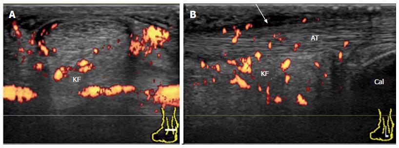
Figure 19 Achilles paratenonitis in early rheumatoid arthritis.
Transverse (A) and longitudinal (B) power Doppler sonogram of the AT shows thickened, hypoechoic, and hyperemic paratenon (arrow) at the level of the KF with minimal tendinopathy. Hyperemia in the KF is also depicted. AT: Achilles tendon; KF: Kager’s fat pad; Cal: Calcaneus.
- Citation: Suzuki T. Power Doppler ultrasonographic assessment of the ankle in patients with inflammatory rheumatic diseases. World J Orthop 2014; 5(5): 574-584
- URL: https://www.wjgnet.com/2218-5836/full/v5/i5/574.htm
- DOI: https://dx.doi.org/10.5312/wjo.v5.i5.574


