Copyright
©2014 Baishideng Publishing Group Inc.
World J Orthop. Nov 18, 2014; 5(5): 574-584
Published online Nov 18, 2014. doi: 10.5312/wjo.v5.i5.574
Published online Nov 18, 2014. doi: 10.5312/wjo.v5.i5.574
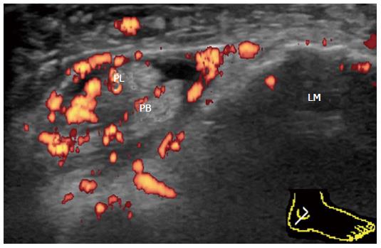
Figure 13 Proliferative tenosynovitis of the peroneus longus and brevis in rheumatoid arthritis.
Transverse power Doppler sonogram through the lateral ankle at the level of the lateral malleolus shows thickening and marked hyperemia of the tenosynovium surrounding the peroneus longus and peroneus brevis. PL: Peroneus longus; PB: Peroneus brevis; LM: Lateral malleolus.
- Citation: Suzuki T. Power Doppler ultrasonographic assessment of the ankle in patients with inflammatory rheumatic diseases. World J Orthop 2014; 5(5): 574-584
- URL: https://www.wjgnet.com/2218-5836/full/v5/i5/574.htm
- DOI: https://dx.doi.org/10.5312/wjo.v5.i5.574


