Copyright
©2014 Baishideng Publishing Group Inc.
World J Orthop. Nov 18, 2014; 5(5): 574-584
Published online Nov 18, 2014. doi: 10.5312/wjo.v5.i5.574
Published online Nov 18, 2014. doi: 10.5312/wjo.v5.i5.574
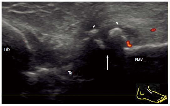
Figure 8 Deformity of the talonavicular joint in rheumatoid arthritis.
Longitudinal power Doppler sonogram of the dorsal aspect of the TNJ shows joint space narrowing (arrow) and osteophyte formation (arrowhead) with minimum flow signal adjacent to an osteophyte. TNJ: Talonavicular joint; Tib: Tibia; Tal: Talus; Nav: Navicular.
- Citation: Suzuki T. Power Doppler ultrasonographic assessment of the ankle in patients with inflammatory rheumatic diseases. World J Orthop 2014; 5(5): 574-584
- URL: https://www.wjgnet.com/2218-5836/full/v5/i5/574.htm
- DOI: https://dx.doi.org/10.5312/wjo.v5.i5.574


