Copyright
©2014 Baishideng Publishing Group Inc.
World J Orthop. Nov 18, 2014; 5(5): 574-584
Published online Nov 18, 2014. doi: 10.5312/wjo.v5.i5.574
Published online Nov 18, 2014. doi: 10.5312/wjo.v5.i5.574
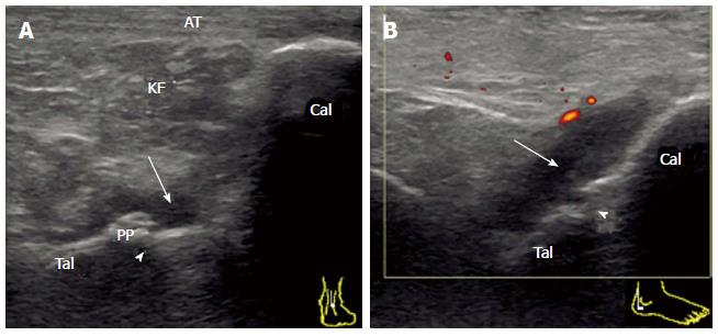
Figure 4 Synovitis of the posterior subtalar joint in rheumatoid arthritis.
A: Sagittal grey-scale sonogram crossing the posterior talar process shows the edge of the distended posterior recess (arrow) of the PSTJ. B: Posterolateral view of the power Doppler sonogram fully depicting the distended posterior recess (arrow) of the PSTJ. Note the close relation of this recess with the superior margin of the calcaneus (Cal). Tib: Tibia; Tal: Talus; KF: Kager’s fat pad; AT: Achilles tendon, arrowhead (joint space); PSTJ: Posterior subtalar joint; PP: Posterior talar process.
- Citation: Suzuki T. Power Doppler ultrasonographic assessment of the ankle in patients with inflammatory rheumatic diseases. World J Orthop 2014; 5(5): 574-584
- URL: https://www.wjgnet.com/2218-5836/full/v5/i5/574.htm
- DOI: https://dx.doi.org/10.5312/wjo.v5.i5.574


