Copyright
©2014 Baishideng Publishing Group Inc.
World J Orthop. Jul 18, 2014; 5(3): 272-282
Published online Jul 18, 2014. doi: 10.5312/wjo.v5.i3.272
Published online Jul 18, 2014. doi: 10.5312/wjo.v5.i3.272
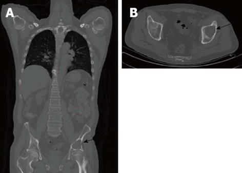
Figure 2 Computed tomography of an osseous myeloma lesion.
Computed tomography in coronal (A) and transversal views (B) showing an osteolytic lesion in the left iliac bone (arrows) representing an osseous myeloma manifestation.
- Citation: Derlin T, Bannas P. Imaging of multiple myeloma: Current concepts. World J Orthop 2014; 5(3): 272-282
- URL: https://www.wjgnet.com/2218-5836/full/v5/i3/272.htm
- DOI: https://dx.doi.org/10.5312/wjo.v5.i3.272


