
















©2014 Baishideng Publishing Group Co.
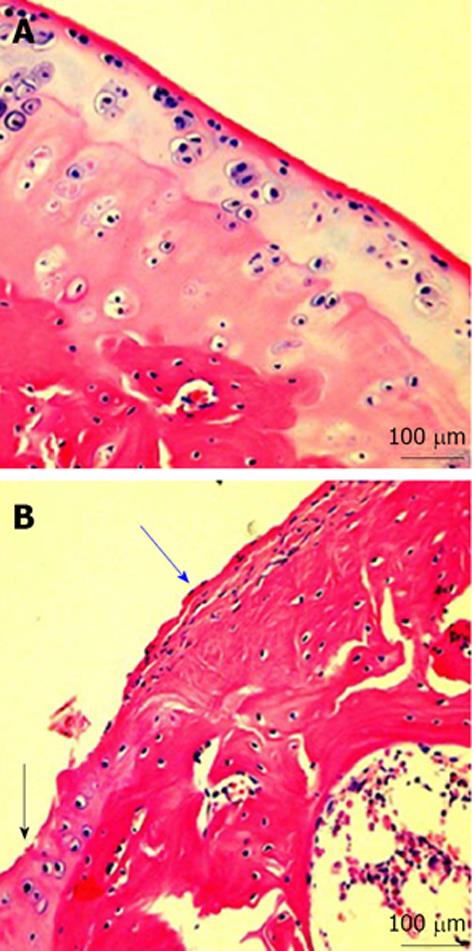
Figure 2 Microscopic signs.
A: Microscopic signs of healthy knee hyaline cartilage. The histological (HE staining) analysis of cartilage from normal donor, showed a preserved morphological structure with no sign of cartilage degradation. Moreover, the surface of healthy hyaline cartilage appears white, shiny, elastic and firm. Magnification x 20; Scale bars: 100 μm; B: Microscopic signs of osteoarthritis (OA) knee hyaline cartilage. The histological (HE staining) analysis of cartilage from OA donor. The donor demonstrated joint swelling and oedema, horizontal cleavage tears or flaps, the surface becomes dull and irregular and had minimal healing capacity. Magnification x 20; Scale bars: 100 μm. Moderate OA cartilage (black arrow), the structural alterations included a reduction of cartilage thickness of the superficial and the middle zones. The structure of the collagen network is damaged, which leads to reduced thickness of the cartilage. The chondrocytes are unable to maintain their repair activity with subsequent loss of the cartilage tissue. Severe OA cartilage (blue arrow), demonstrated deep surface clefts, disappearance of cells from the tangential zone, cloning, and a lack of cells in the intermediate and radial zone, which are not arranged in columns. The tidemark is no longer intact and the subchondral bone shows fibrillation.
- Citation: Musumeci G, Castrogiovanni P, Leonardi R, Trovato FM, Szychlinska MA, Di Giunta A, Loreto C, Castorina S. New perspectives for articular cartilage repair treatment through tissue engineering: A contemporary review. World J Orthop 2014; 5(2): 80-88
- URL: https://www.wjgnet.com/2218-5836/full/v5/i2/80.htm
- DOI: https://dx.doi.org/10.5312/wjo.v5.i2.80