Copyright
©2013 Baishideng Publishing Group Co.
World J Orthop. Jul 18, 2013; 4(3): 154-156
Published online Jul 18, 2013. doi: 10.5312/wjo.v4.i3.154
Published online Jul 18, 2013. doi: 10.5312/wjo.v4.i3.154
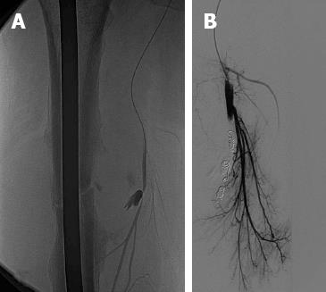
Figure 3 Embolisation.
Elective (microcatheter) right deep femoral angiography pictures showing the pseudoaneurysm adjacent to the fracture site. A: Before coil embolization; B: After coil embolization (digital subtraction angiography).
- Citation: Valli F, Teli MG, Innocenti M, Vercelli R, Prestamburgo D. Profunda femoris artery pseudoaneurysm following revision for femoral shaft fracture nonunion. World J Orthop 2013; 4(3): 154-156
- URL: https://www.wjgnet.com/2218-5836/full/v4/i3/154.htm
- DOI: https://dx.doi.org/10.5312/wjo.v4.i3.154


