Copyright
©2013 Baishideng Publishing Group Co.
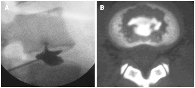
Figure 2 Discography and computed tomography.
A: Discography showing a radial disruption on the lower endplate of L4 vertebra and that the contrast medium flows into the cancellous bone of the lower endplate of L4 vertebra through the fissure; B: Computed tomography scan showing the contrast medium dispersed in the lower endplate of L4 vertebra, with Grade 4 endplate disruption.
- Citation: Peng BG. Pathophysiology, diagnosis, and treatment of discogenic low back pain. World J Orthop 2013; 4(2): 42-52
- URL: https://www.wjgnet.com/2218-5836/full/v4/i2/42.htm
- DOI: https://dx.doi.org/10.5312/wjo.v4.i2.42


