Copyright
©2011 Baishideng Publishing Group Co.
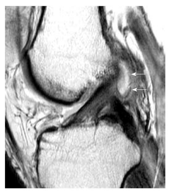
Figure 20 Pericruciate ganglion cyst.
Sagittal intermediate-weighted MR image shows a ganglion cyst (white arrows) arising from proximal posterior half of ligament.
- Citation: Ng WHA, Griffith JF, Hung EHY, Paunipagar B, Law BKY, Yung PSH. Imaging of the anterior cruciate ligament. World J Orthop 2011; 2(8): 75-84
- URL: https://www.wjgnet.com/2218-5836/full/v2/i8/75.htm
- DOI: https://dx.doi.org/10.5312/wjo.v2.i8.75


