Copyright
©2011 Baishideng Publishing Group Co.
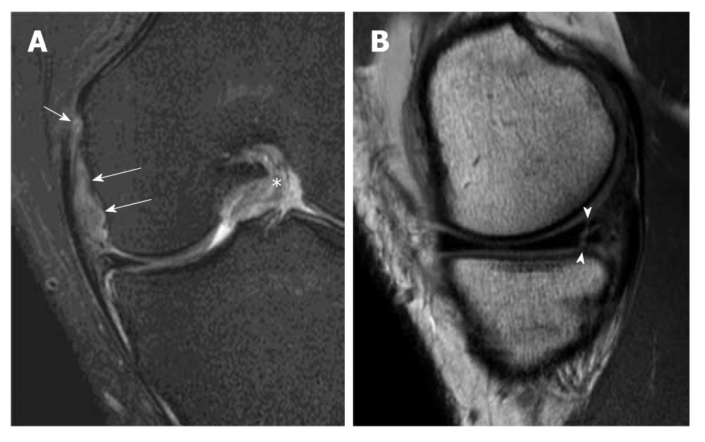
Figure 17 O’ Donoghue’s triad.
Magnetic resonance images of a patient with complete anterior cruciate ligament (ACL) tear showing O’ Donoghue’s triad. A: Coronal T2-weighted fat suppression magnetic resonance (MR) image demonstrates complete tear of the meniscofemoral ligament (white long arrows) and femoral attachment of the medial collateral ligament (white short arrow). Note that the ACL in the intercondylar fossa has a substantial tear (asterisk); B: Sagittal intermediate-weighted MR image shows an associated vertical and horizontal (complex) peripheral tear at the posterior horn of medial meniscus (arrowheads). These injuries constitute the classical “O Donoghue’s triad”.
- Citation: Ng WHA, Griffith JF, Hung EHY, Paunipagar B, Law BKY, Yung PSH. Imaging of the anterior cruciate ligament. World J Orthop 2011; 2(8): 75-84
- URL: https://www.wjgnet.com/2218-5836/full/v2/i8/75.htm
- DOI: https://dx.doi.org/10.5312/wjo.v2.i8.75


