Copyright
©2011 Baishideng Publishing Group Co.
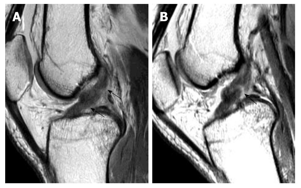
Figure 9 Magnetic resonance knee in partial flexion.
Volunteer, 31-year-old man with no history of injury and clinical instability. Sagittal intermediate-weighted magnetic resonance image in full extension. A: And 30 degree of knee flexion; B: Demonstrates the usefulness of knee flexion. When the knee is extended, sagittal image shows features suspicious of an anterior cruciate ligament (ACL) tear. When the knee is flexed, a gap (black arrow) is clearly present confirming the presence of an ACL tear.
- Citation: Ng WHA, Griffith JF, Hung EHY, Paunipagar B, Law BKY, Yung PSH. Imaging of the anterior cruciate ligament. World J Orthop 2011; 2(8): 75-84
- URL: https://www.wjgnet.com/2218-5836/full/v2/i8/75.htm
- DOI: https://dx.doi.org/10.5312/wjo.v2.i8.75


