Copyright
©2011 Baishideng Publishing Group Co.
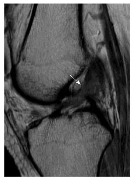
Figure 8 Partial tear of the anterior cruciate ligament.
Sagittal intermediate-weighted image of partial anterior cruciate ligament (ACL) tear. The ACL appears lax, concave in appearance (white arrow) and increased in signal intensity. However, the fibres are still in continuity suggestive of partial ACL tear.
- Citation: Ng WHA, Griffith JF, Hung EHY, Paunipagar B, Law BKY, Yung PSH. Imaging of the anterior cruciate ligament. World J Orthop 2011; 2(8): 75-84
- URL: https://www.wjgnet.com/2218-5836/full/v2/i8/75.htm
- DOI: https://dx.doi.org/10.5312/wjo.v2.i8.75


