Copyright
©2011 Baishideng Publishing Group Co.
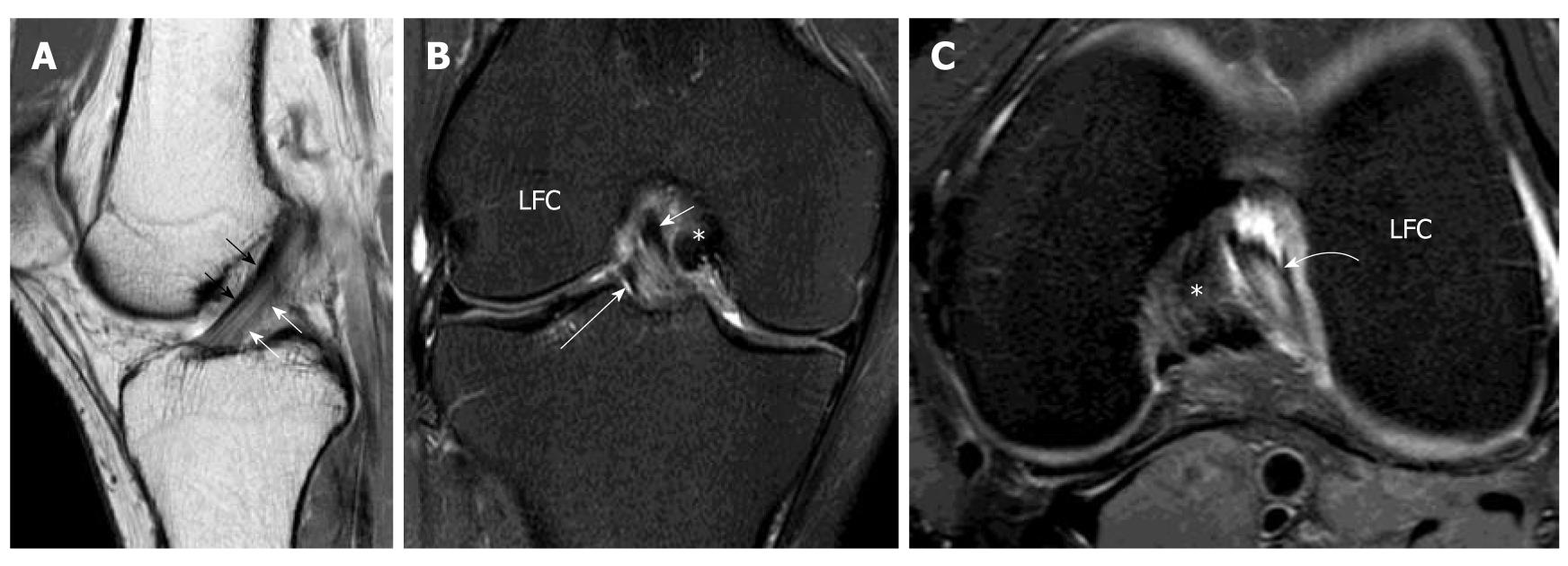
Figure 4 Normal anterior cruciate ligament.
Volunteer, 24-year-old man with no history of injury and clinical instability. Sagittal intermediate-weighted magnetic resonance (MR) of the knee. A: Demonstrates a normal anterior cruciate ligament (ACL) which is characterized by a taut, continuous, low signal intensity fibres extending from the tibial plateau anteriorly to the medial aspect of the lateral femoral condyle. The more anterior portion of ACL is the anteromedial (AM) bundle (black arrows) while the more posterior portion is posterolateral (PL) bundle (white arrows). They cannot be well delineated from each other on this sagittal image. The PL bundle shows higher signal intensity than AM bundle. Coronal T2-weighted fat suppression MR knee image; B: The mid and distal ACL in the intercondylar fossa. The fibres are running superiorly and laterally within the intercondylar fossa from tibial attachment to the lateral femoral condyle (LFC). The more medial portion is the AM bundle (white short arrow) while the lateral portion is the PL bundle (white long arrow). Axial intermediate-weighted fat suppression MR image; C: Normal mid substance of ACL (curved white arrow). The ACL is elliptical in appearance because it is running obliquely to the scan plane. The two bundles cannot be differentiated from each other. *: Posterior cruciate ligament.
- Citation: Ng WHA, Griffith JF, Hung EHY, Paunipagar B, Law BKY, Yung PSH. Imaging of the anterior cruciate ligament. World J Orthop 2011; 2(8): 75-84
- URL: https://www.wjgnet.com/2218-5836/full/v2/i8/75.htm
- DOI: https://dx.doi.org/10.5312/wjo.v2.i8.75


