
















Copyright: ©Author(s) 2026.
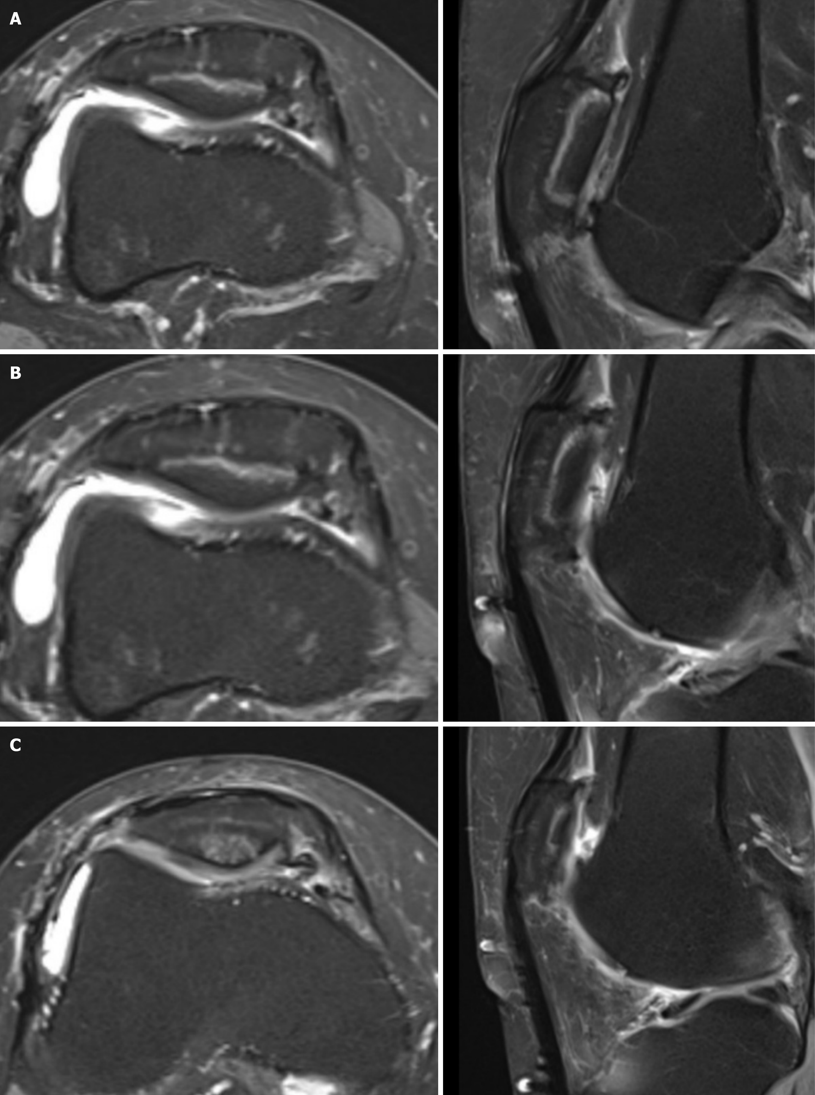
Figure 3 Non-contrast magnetic resonance.
A-C: Showing sagittal and axial T2 slices of the patella 32 months post-operation demonstrating a retained osteochondral allograft with maintained articular congruity and intact subchondral bone bed. With overall improved fluid accumulation at the base of the allograft.
- Citation: Desai B, Willard JG, Assid E, Hall C, Renshaw A, Schoofs E, Wilder JH, Hatcher L, Tupler R, Jones D. Missouri osteochondral preservation system transplantation: Outcome scores from 24 months to 60+ months. World J Orthop 2026; 17(9): 114039
- URL: https://www.wjgnet.com/2218-5836/full/v17/i9/114039.htm
- DOI: https://dx.doi.org/10.5312/wjo.114039