
















Copyright: ©Author(s) 2026.
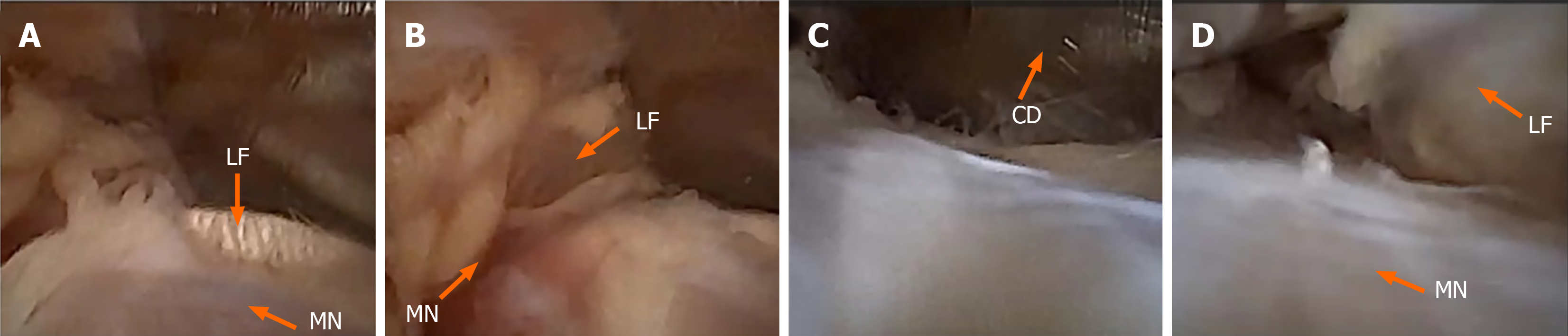
Figure 2 Sequential nanoscopic views during nanoscope-assisted lacertus fibrosus release.
A: Identification of the lacertus fibrosus and underlying median nerve; B: Development of the working corridor with visualization of the lacertus fibrosus and median nerve; C: Introduction of the cutting device under direct visualization; D: Final nanoscopic view following controlled division of the lacertus fibrosus, demonstrating the underlying median nerve. LF: Lacertus fibrosus; MN: Median nerve; CD: Cutting device.
- Citation: Chlebanowski B, Wojtowicz BG, Urban K, Kanak M, Domzalski M, Lesman J. Nanoscope-assisted release of the lacertus fibrosus: A cadaveric study of feasibility, visualization, and anatomical safety. World J Orthop 2026; 17(8): 123794
- URL: https://www.wjgnet.com/2218-5836/full/v17/i8/123794.htm
- DOI: https://dx.doi.org/10.5312/wjo.123794