
















Copyright: ©Author(s) 2026.
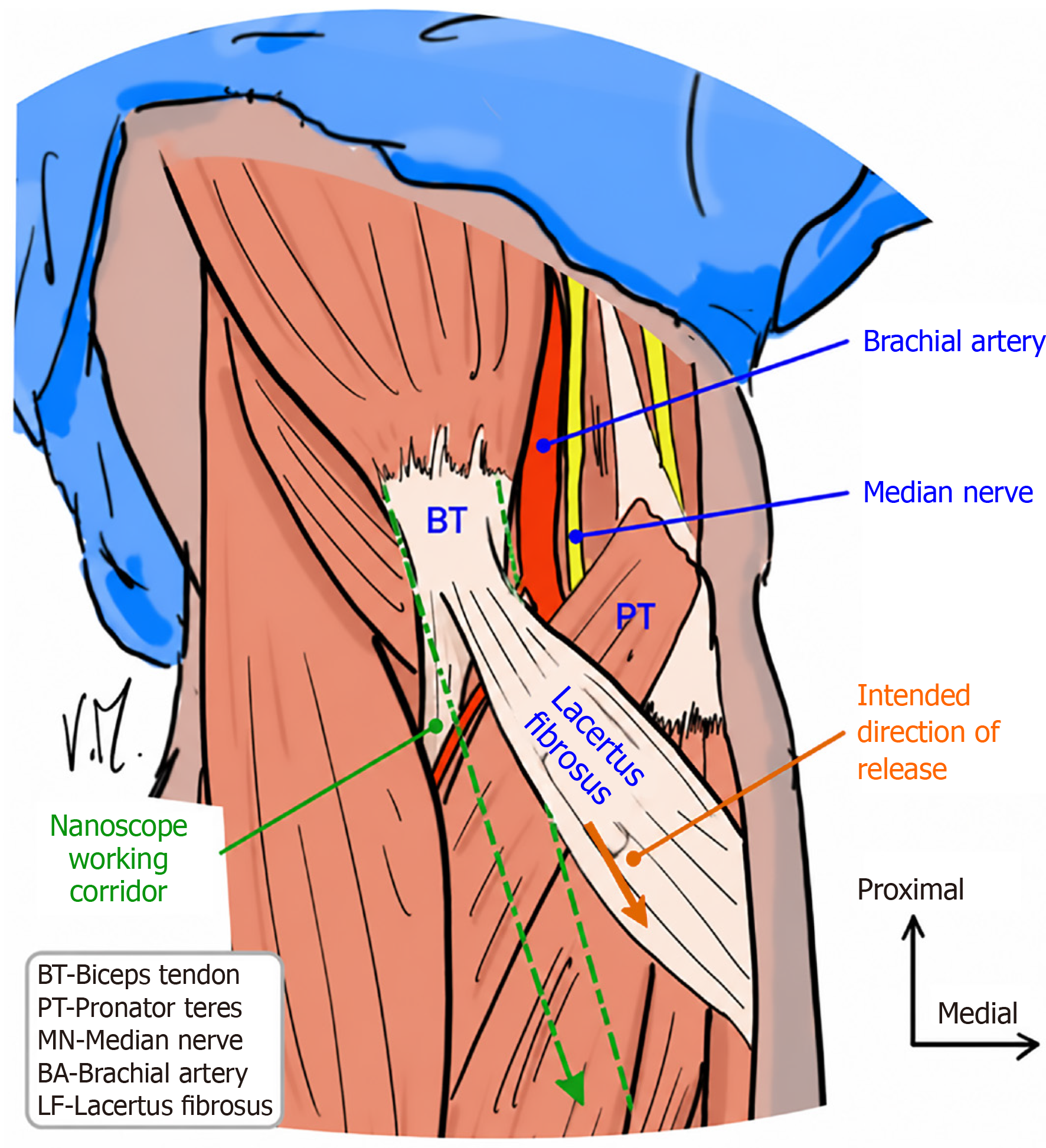
Figure 1 Schematic illustration of the proximal forearm anatomy demonstrating the anatomical relationship between the lacertus fibrosus, median nerve, brachial artery, and pronator teres.
The lacertus fibrosus is shown crossing superficially over the median nerve and adjacent vascular structures. BT: Biceps tendon; PT: Pronator teres; MN: Median nerve; BA: Brachial artery; LF: Lacertus fibrosus.
- Citation: Chlebanowski B, Wojtowicz BG, Urban K, Kanak M, Domzalski M, Lesman J. Nanoscope-assisted release of the lacertus fibrosus: A cadaveric study of feasibility, visualization, and anatomical safety. World J Orthop 2026; 17(8): 123794
- URL: https://www.wjgnet.com/2218-5836/full/v17/i8/123794.htm
- DOI: https://dx.doi.org/10.5312/wjo.123794