
















Copyright: ©Author(s) 2026.
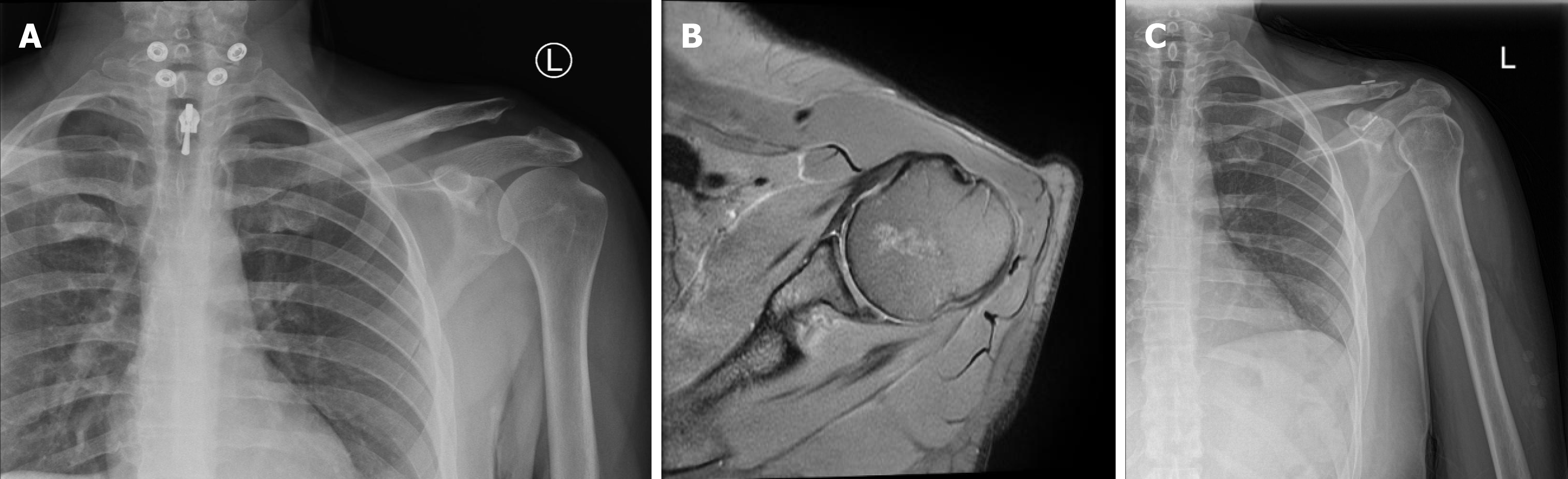
Figure 1 Diagnostic imaging.
A: X-ray showing acromioclavicular joint dislocation; B: Magnetic resonance imaging image showing the soft tissue Bankart lesion of the anterior glenoid and the Hill-Sachs lesion of the posterior humeral head; C: X-ray showing the acromioclavicular joint postoperatively.
- Citation: Al Hulaibi FH, AlBassam AY, AlGhamdi OS, Alahmari AS, Altwejry AY, Almatrouk JM, Alzaid ZA. Dual arthroscopic approach for simultaneous Bankart lesion repair and acromioclavicular joint stabilization: A case report. World J Orthop 2026; 17(8): 122866
- URL: https://www.wjgnet.com/2218-5836/full/v17/i8/122866.htm
- DOI: https://dx.doi.org/10.5312/wjo.122866