
















Copyright: ©Author(s) 2026.
World J Orthop. Jun 18, 2026; 17(6): 121411
Published online Jun 18, 2026. doi: 10.5312/wjo.v17.i6.121411
Published online Jun 18, 2026. doi: 10.5312/wjo.v17.i6.121411
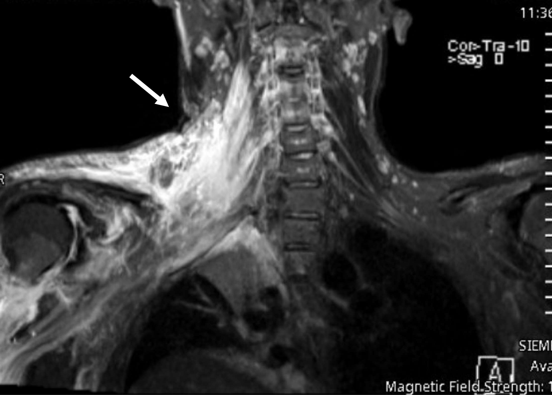
Figure 4 Magnetic resonance imaging shows brachial plexus with evidence of significant subcutaneous oedema noted along the right lower cervical and shoulder regions with irregular soft tissue hematoma (arrow) along the inferior cervical region extending into the right supraclavicular and right axillary region displacing and partially entrapping the trunks of ipsilateral brachial plexus which appears relatively thickened with abnormal high T2WI signal intensity.
Underlying brachial plexus injury cannot be excluded and might be masked by the soft tissue hematoma.
- Citation: Al-Thani H, El-Menyar A, Asim M, Syed Y, Ghouri SI, Muneer M, Al-Hassani A, Al-Sulaiti M, Tabeb A. Management and outcome of blunt traumatic brachial plexus injuries in adult trauma patients: A retrospective study. World J Orthop 2026; 17(6): 121411
- URL: https://www.wjgnet.com/2218-5836/full/v17/i6/121411.htm
- DOI: https://dx.doi.org/10.5312/wjo.v17.i6.121411