
















Copyright: ©Author(s) 2026.
World J Orthop. Jun 18, 2026; 17(6): 121411
Published online Jun 18, 2026. doi: 10.5312/wjo.v17.i6.121411
Published online Jun 18, 2026. doi: 10.5312/wjo.v17.i6.121411
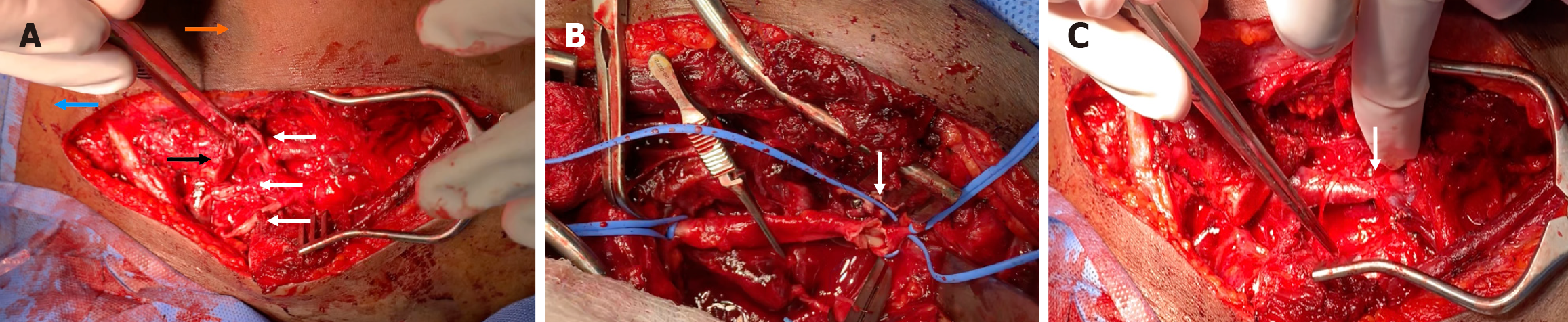
Figure 3 The examination results during the surgical procedure.
A: Orange arrow, pointing to the neck of the patient, the blue arrow pointing to the right shoulder, the black arrow pointing to the right clavicle, which is divided to expose the neurovascular of the brachial plexus and the axillary artery and vein. The three white arrows show the complete transaction of the nerve at the level of the cords of the brachial plexus; B: Proximal auxiliary artery with the repair process and vascular clamp on the proximal and distal ends for control; C: Axillary vein after repair.
- Citation: Al-Thani H, El-Menyar A, Asim M, Syed Y, Ghouri SI, Muneer M, Al-Hassani A, Al-Sulaiti M, Tabeb A. Management and outcome of blunt traumatic brachial plexus injuries in adult trauma patients: A retrospective study. World J Orthop 2026; 17(6): 121411
- URL: https://www.wjgnet.com/2218-5836/full/v17/i6/121411.htm
- DOI: https://dx.doi.org/10.5312/wjo.v17.i6.121411