
















Copyright: ©Author(s) 2026.
World J Orthop. Jun 18, 2026; 17(6): 121048
Published online Jun 18, 2026. doi: 10.5312/wjo.v17.i6.121048
Published online Jun 18, 2026. doi: 10.5312/wjo.v17.i6.121048
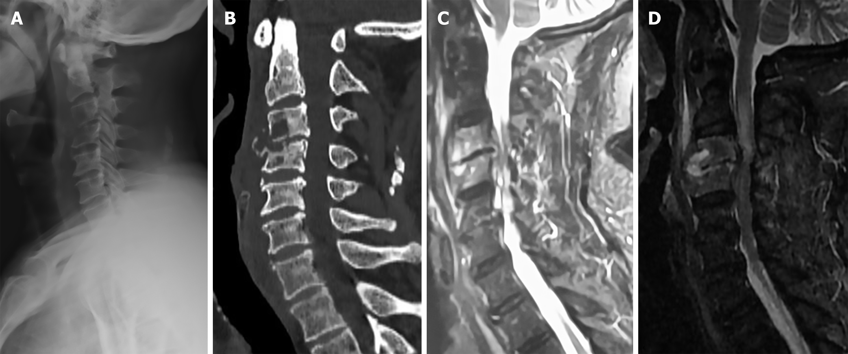
Figure 1 Imaging findings on admission and before surgery.
A: X-ray imaging upon admission revealing low-density shadows along the front of the C3 and C4 vertebrae, with an interruption of the bone cortex and a decrease in the C3/4 intervertebral space; B: Computed tomography scan upon admission showing destruction of the bone within the C3 and C4 vertebrae; C: Admission MRI revealing hyperintensity on T2-weighted fat-suppressed images at the anterior margins of the C3 and C4 vertebral bodies, with mild spinal cord compression; D: Preoperative MRI demonstrating hyperintensity on T2-weighted fat-suppressed images involving the C3 and C4 vertebral bodies and intervertebral space, with marked spinal cord compression.
- Citation: Wang S, Ren SW, Chang ZQ, Li CH. Utilization of autogenous iliac bone shaped into a low-profile form to repair bone defects following cervical infection: A case report. World J Orthop 2026; 17(6): 121048
- URL: https://www.wjgnet.com/2218-5836/full/v17/i6/121048.htm
- DOI: https://dx.doi.org/10.5312/wjo.v17.i6.121048